The analysis
Six parameters. One objective picture of the scalp.
Every Trichora scan returns the same structured set of findings, so two visits — or two clinics — are always comparing like with like.
Parameter 01
Hairline recession map
From the front view, Trichora traces the current hairline and compares it with the expected position for the patient's facial proportions. The gap between the two — across the mid-forehead and both temple corners — is drawn as a contour the patient can see at a glance.
- Output
Annotated front image with expected vs. actual hairline
- Why it matters
Temple recession is the earliest pattern most patients notice last

Parameter 02
Baldness grade, 1 to 7
The hairline map, temple-corner recession and crown involvement combine into a single clinical staging grade every dermatologist already speaks. Select a stage to see the scalp and what Trichora reports for it.
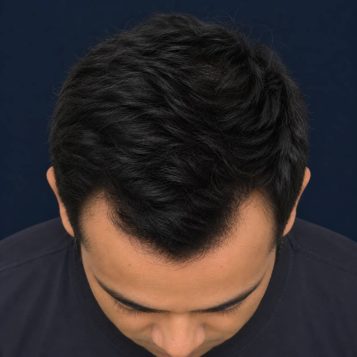
Female pattern · Ludwig I–III
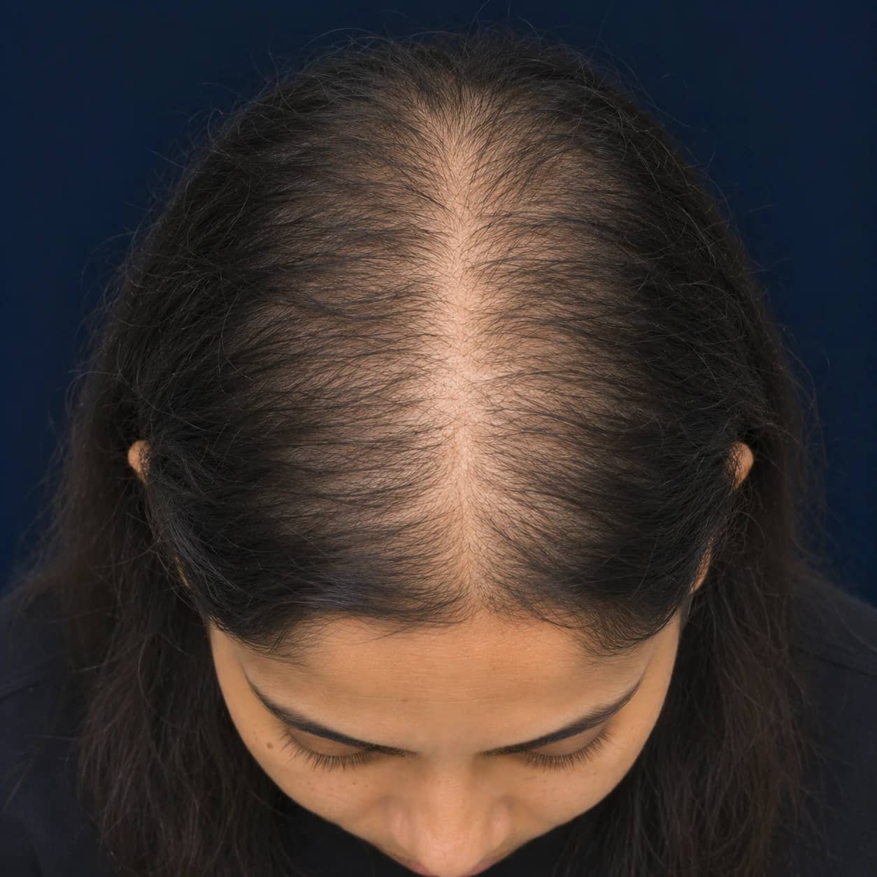
Women are staged on their own scale.
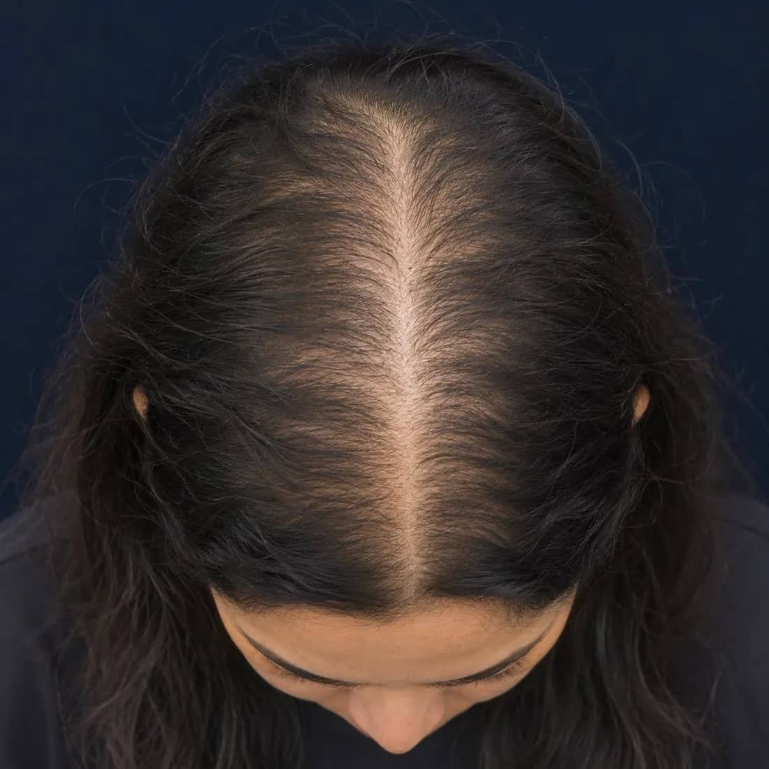
Female-pattern loss keeps the hairline and widens the parting, so a 1–7 recession grade would mis-stage every woman. Trichora reports women on the three-stage Ludwig scale from the same top view.
Parameters 03 & 04
Zone thinning, scored 0–100%
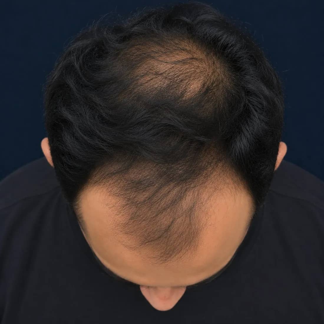
From the top view, the scalp is read as three zones — frontal, mid-scalp and crown. In each, Trichora outlines the region of density loss and scores how much scalp shows through, from 0% (full coverage) to 100% (fully exposed).
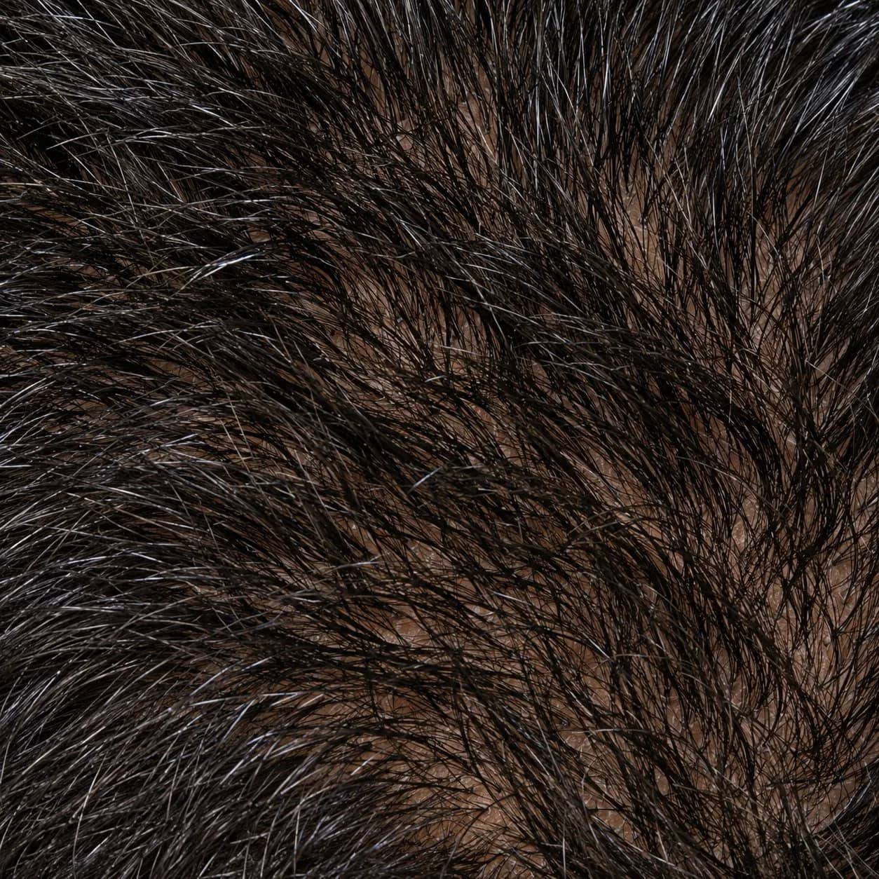 0%Full densityNo scalp visible through hair
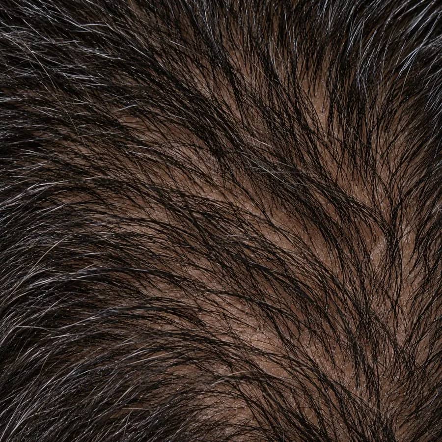 25%Early thinningReduced density; scalp not yet visible
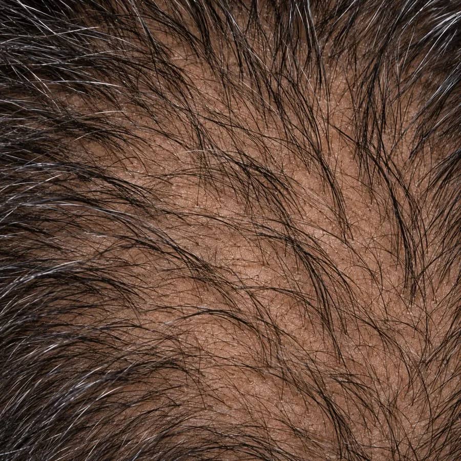 50%ModerateScalp partially visible
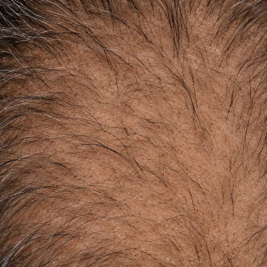 75%AdvancedScalp clearly visible
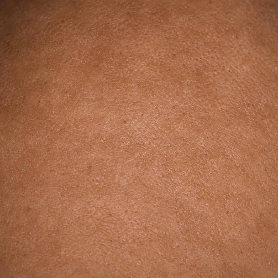 100%Fully exposedNo remaining coverage in the zone
- Frontal
The band behind the hairline. Often the first zone to thin and the one patients notice in the mirror.
- Mid-scalp
The bridge between front and crown. Scored independently so a healthy bridge doesn't hide what's happening behind it.
- Crown
The vertex. Invisible to the patient, visible to everyone else — and the zone most often under-treated.
Parameters 05 & 06
Temporal and occipital flags
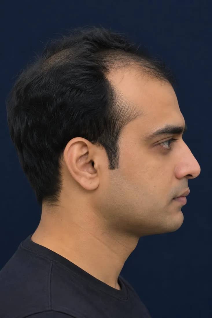
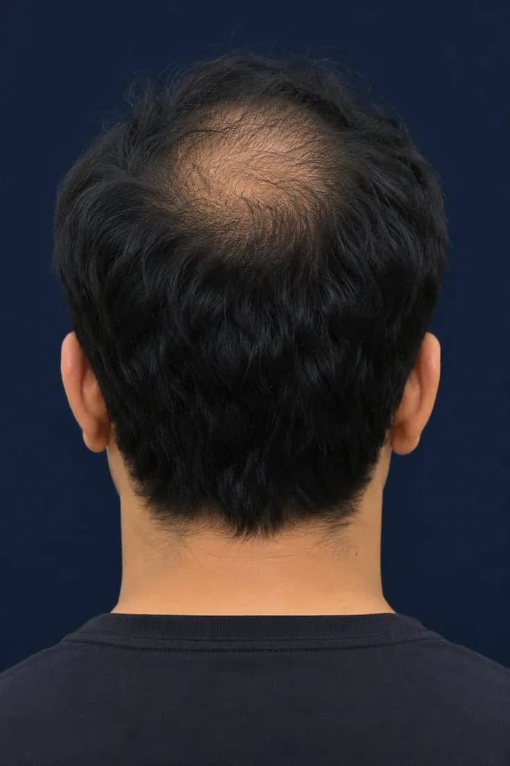
From the left, right and back views, Trichora checks the band above each ear and the nape and lower crown for any thinning. These regions are reported as present or absent rather than scored — a clear early-warning signal and a check on donor-area integrity before any transplant conversation.
- Temporal leftClear
- Temporal rightThinning present
- OccipitalClear
- Left· clear
- Right· present

- Back· clear
What every report contains
Hairline map
- Source view
- Front
- Output
- Contour · expected vs actual
- Used for
- Patient explanation, grade
Baldness grade
- Source view
- Front + top
- Output
- 1–7
- Used for
- Staging, progression outlook
Frontal thinning
- Source view
- Top
- Output
- Contour · 0–100%
- Used for
- Protocol, tracking
Mid-scalp & crown thinning
- Source view
- Top
- Output
- Contour · 0–100% each
- Used for
- Protocol, tracking
Temporal flags
- Source view
- Left, right
- Output
- Present / absent
- Used for
- Early warning
Occipital flags
- Source view
- Back
- Output
- Present / absent
- Used for
- Donor-area check
| Parameter | Source view | Output | Used for |
|---|---|---|---|
| Hairline map | Front | Contour · expected vs actual | Patient explanation, grade |
| Baldness grade | Front + top | 1–7 | Staging, progression outlook |
| Frontal thinning | Top | Contour · 0–100% | Protocol, tracking |
| Mid-scalp & crown thinning | Top | Contour · 0–100% each | Protocol, tracking |
| Temporal flags | Left, right | Present / absent | Early warning |
| Occipital flags | Back | Present / absent | Donor-area check |