For dermatologists, trichologists & hair-restoration surgeons
Objective, repeatable scalp measurement — inside a standard consult.
Five standardised photographs become a Norwood–Hamilton or Ludwig grade, a thinning percentage for each scalp zone and side and occipital flags — drawn as contours on the patient's own images. You see them next to the patient's health-questionnaire answers, review the prescription drafted from your clinic's own protocol, edit and sign. Nothing reaches the patient without you.
- five-view capture
- ~90 s
- clinic report generated
- < 60 s
- doctor review & approval
- ~2 min
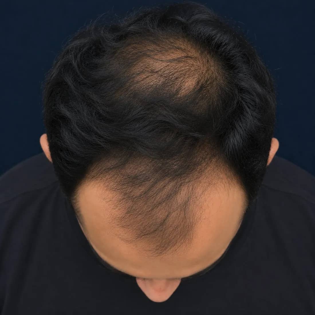
- Grade
- Norwood 4
- Frontal
- 50%
- Mid-scalp
- 25%
- Crown
- 75%
- Flags
- Temporal Rothers clear
What is measured
Five views. Each region read from the angle that shows it.
Every scan returns the same structured set of findings, so a baseline and a follow-up — or two clinicians — are always comparing like with like.
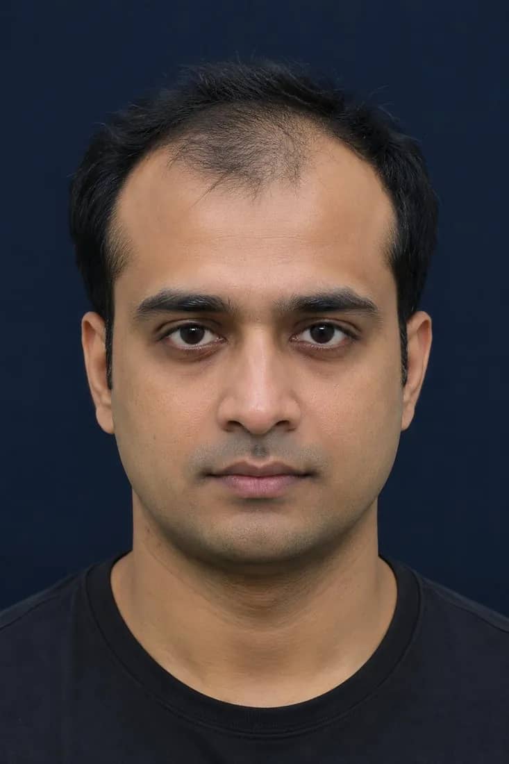
Front
GradeFrontal hairline and temporal recession at both temple corners
- Output
- Hairline contour · contributes to the grade
- Clinical relevance
- Staging. Distinguishes a receding pattern from a preserved hairline — the key separation between male- and female-pattern presentations.
Top
0–100%Frontal zone, mid-scalp and vertex / crown
- Output
- Zone thinning %, 0–100, per zone · contours
- Clinical relevance
- Where to target treatment and how each zone responds. The vertex is the zone the patient cannot see and most often under-reports.
Left & right
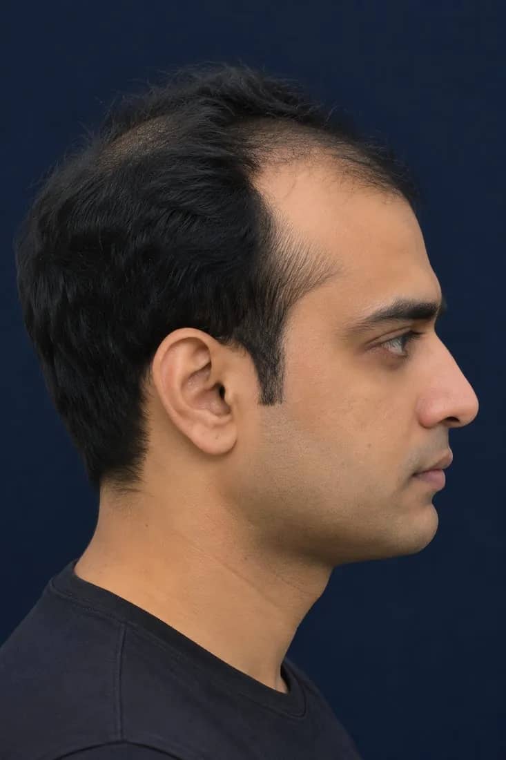
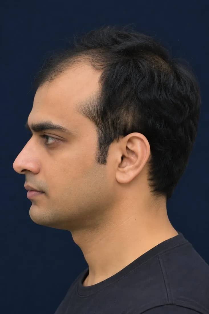
FlagTemporal and parietal bands above each ear
- Output
- Flag · thinning present / absent, each side
- Clinical relevance
- Signals thinning beyond the frontal–vertex territory, and asymmetry between sides, for you to examine.
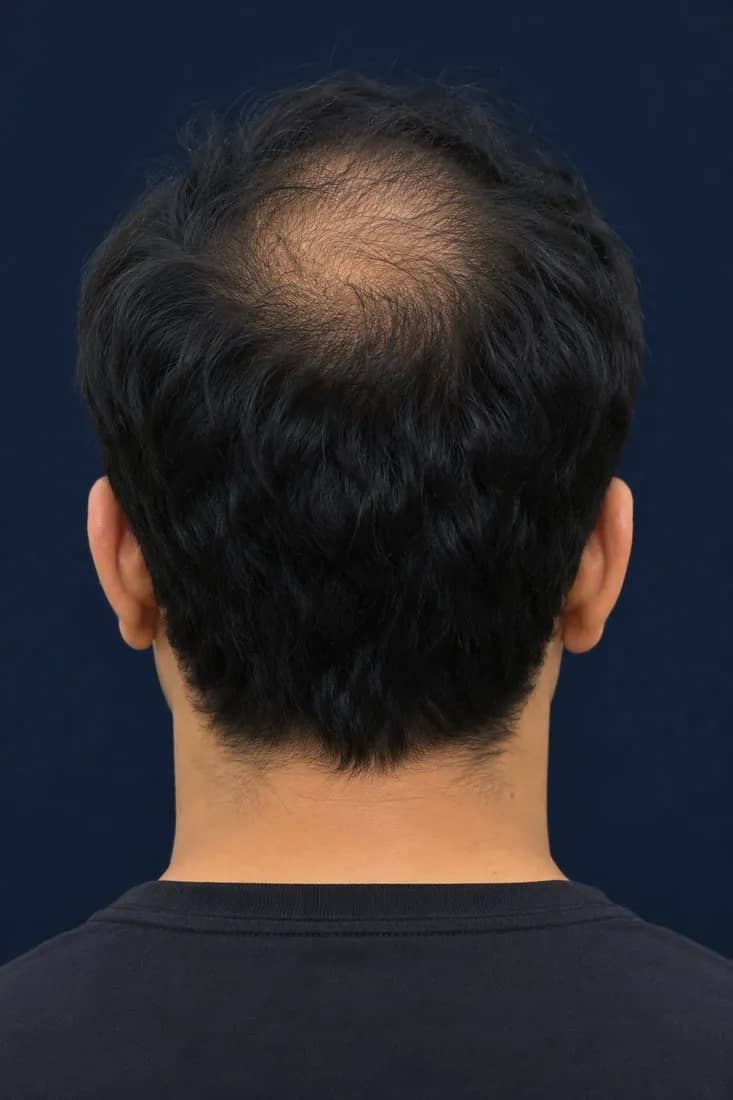
Back
FlagOccipital (donor) area
- Output
- Flag · thinning present / absent
- Clinical relevance
- Useful context when transplant candidacy is being considered. A flag prompts closer clinical assessment of the donor area; it is not a donor-density measurement.
Grade is read from the front and top views together. Side and back regions are reported as flags, not percentages.
Reading the numbers
What “Crown 50%” actually means.
Zone thinning % is the share of scalp visible through the hair within that zone, in the standardised photograph. Crown 50% means half of the scalp surface in the crown zone shows through. 0% is full coverage; 100% is fully exposed scalp.
- Not a hair count
- Not density in hairs/cm²
- Not shaft diameter or miniaturisation
Read each zone against its own baseline: crown against crown, visit against visit. Percentages are comparable because the views, framing and capture checks are the same every time.
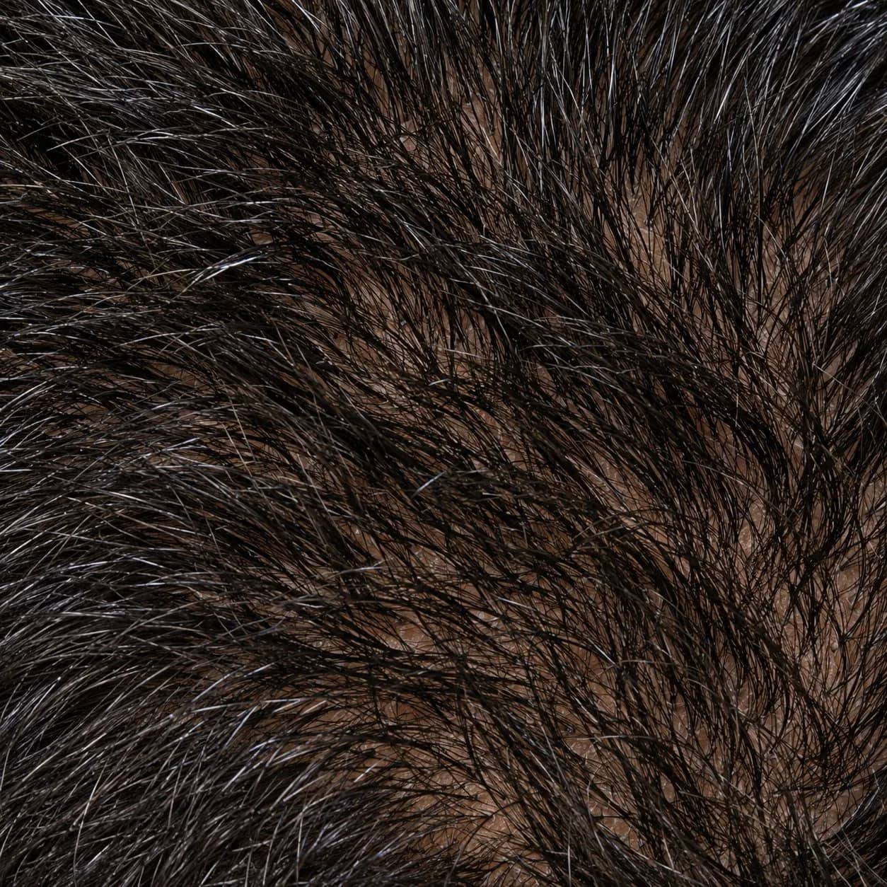 0%Full densityNo scalp visible through hair
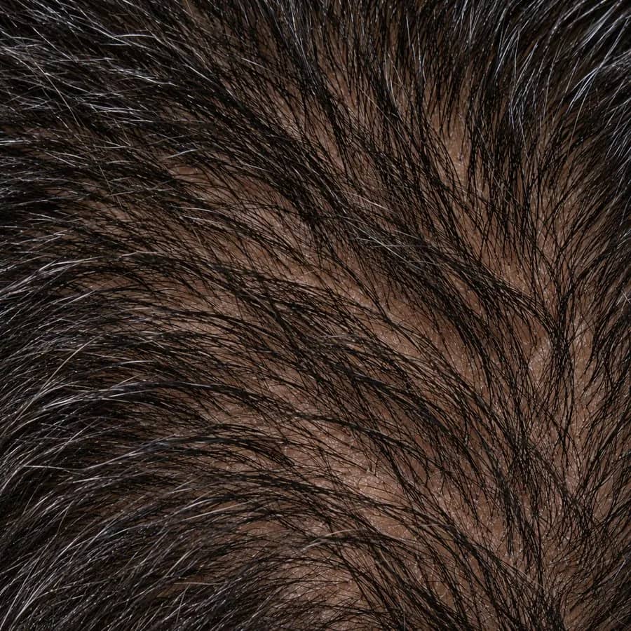 25%Early thinningReduced density; scalp not yet visible
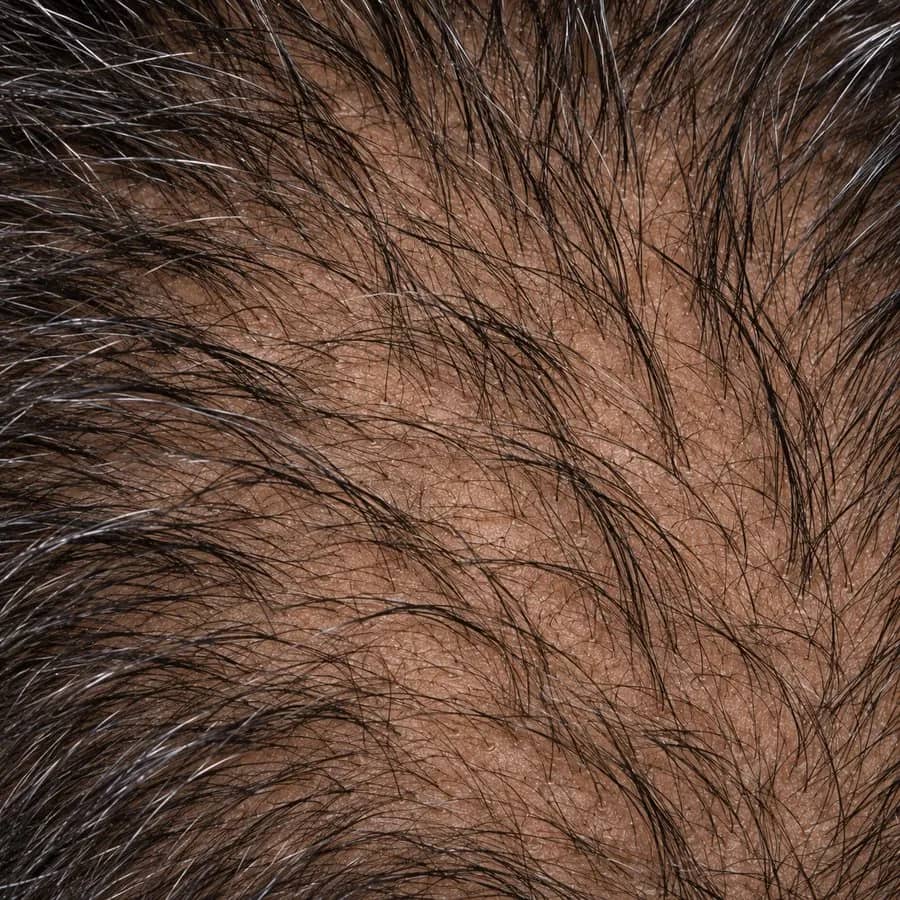 50%ModerateScalp partially visible
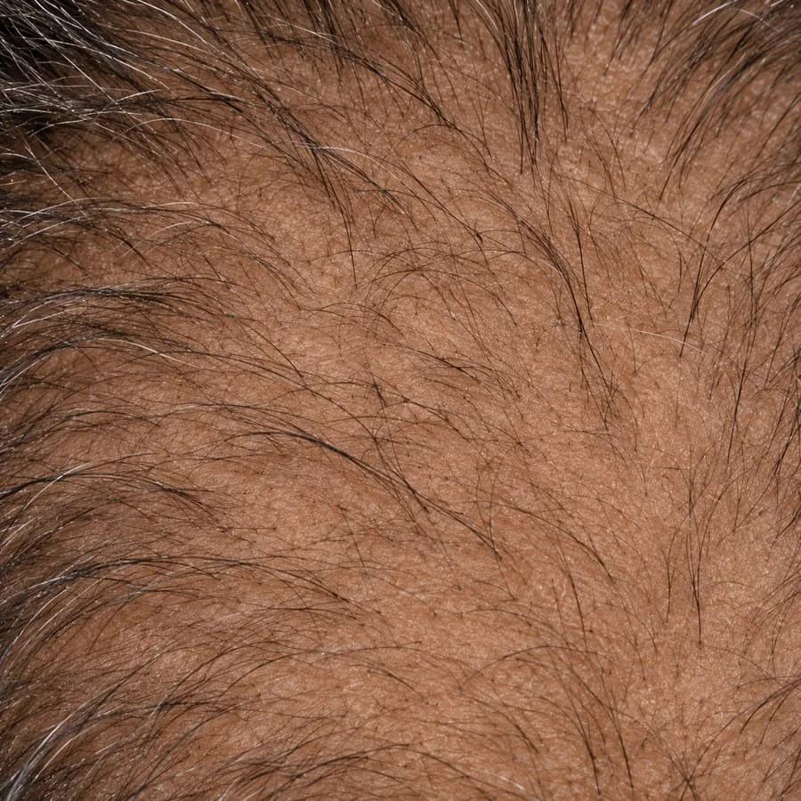 75%AdvancedScalp clearly visible
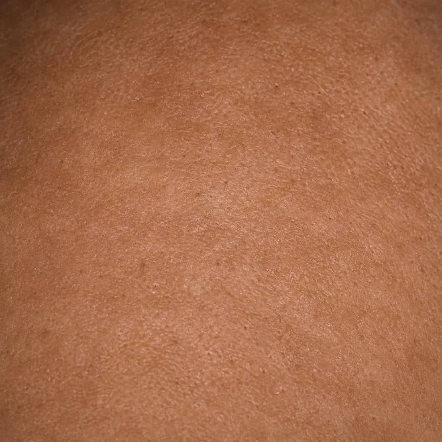 100%Fully exposedNo remaining coverage in the zone
What a scan returns — and what it doesn't
There is no overall score. A single number would hide exactly what you need: which zone, how much, and whether it is changing.
- Grade
- Norwood–Hamilton 1–7 or Ludwig I–III
- Zone thinning %
- Frontal, mid-scalp and crown, each 0–100
- Flags
- Temporal left, temporal right, occipital
Staging
Norwood–Hamilton 1–7 and Ludwig I–III, detected from the images.
The model proposes a stage from the front and top views. Select a stage to see the reference top view and the zone pattern typically reported with it.
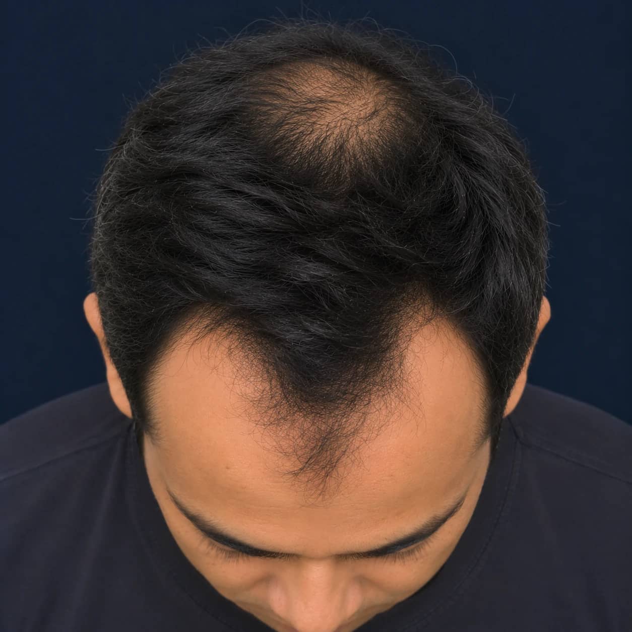 Reference · top view
Deeper frontal recession and a separate round thinning area at the crown. A wide band of full hair still separates them.
Standardised photography
The same five views, every visit. Baselines you can actually compare.
Global photography is only useful for follow-up when it is reproducible. Trichora fixes the views and guides the capture, so a month-4 scan is registered against the baseline zone by zone.
- 01 · Front
- 02 · Left
- 03 · Top
- 04 · Right
- 05 · Back
Fixed views
Front, left, right, top and back — the same set for every patient, in clinic or at home.
Guided alignment
On-screen framing guides head position and distance for each view.
Quality checks
Each photo is checked before analysis, so a poor capture is retaken rather than measured.
Staff or patient
Captured by clinic staff, or uploaded by the patient from the clinic's app.
Change since baseline · month 4
TR-2107 · sample- Frontal50 → 42% ▼
- Mid-scalp25 → 25% =
- Crown75 → 61% ▼
Grade held at 4Faded bar = baseline
Treatment response
Change is reported per zone, so a responding crown and a static mid-scalp are visible separately.
Counselling
Patients see their own annotated photos, not a description — and the 12/24-month outlook, presented as a statistical estimate.
Adherence
Measured change at each re-scan gives patients a reason to stay on a long-horizon protocol.
Where it fits in your assessment
Macroscopic measurement. Not trichoscopy, not a diagnosis.
Trichora standardises and quantifies what global photographs show. It sits alongside the parts of the assessment that need a clinician and a dermatoscope.
Trichora does
- Standardised five-view global photography
- Grade detection: Norwood–Hamilton or Ludwig
- Zone thinning % for frontal, mid-scalp and crown
- Temporal and occipital flags
- Contours on the photos, baseline and follow-up comparison
- A structured health questionnaire, shown next to the photos
- A prescription draft from your clinic's own protocol, for you to edit
You still do
- Confirming and completing the history — the questionnaire is a structured start, not the whole history
- Clinical examination of scalp and hair, and the pull test
- Trichoscopy / dermoscopy
- Investigations, where indicated
- Diagnosis of the cause
- The treatment decision and the prescription
Not measuredHair-shaft diameter, follicular-unit counts, miniaturisation ratio and any perifollicular or scalp-surface sign. These need a dermatoscope and your eyes.
The doctor workflow
From five photos and a short history to a signed prescription — in minutes.
The model detects and measures; your clinic's protocol drafts. You review, edit, approve and sign — about two minutes. Every step is logged.
All 9 steps
- 01Guided photos. Five standard views, captured by staff in clinic or by the patient from the clinic's app, with alignment and quality checks on capture.
- 02Health questionnaire. 10–15 short questions — sex and age, family planning, recent illness or viral infection, duration of loss, and more. You see the answers next to the photos.
- 03AI grade & zone analysis. Norwood–Hamilton or Ludwig grade, thinning % for each zone and side and occipital flags, drawn as contours on the patient's own photos.
- 04Clinic report. The clinic report is generated automatically and the case lands in your queue: annotated views, findings and the questionnaire answers on one screen.
- 05Prescription drafted. Your clinic's own treatment protocol — a fixed rule set your clinic defines — is applied to the report and the answers. The result is a draft, not a prescription.
- 06Doctor approves. Check the findings against the history, add, remove or change any line, then approve. Your name, qualification, registration number and timestamp are applied when you e-sign.
- 07Sent on WhatsApp. The moment you sign, the report, the signed prescription PDF on the clinic letterhead and an order link reach the patient through the official WhatsApp Business Platform, with consent.
- 08Order & doorstep delivery. The link opens the clinic's own order page. A licensed pharmacy partner dispenses against your signed prescription and delivers to the patient's door.
- 09Follow-up & re-scan. Reminders support adherence, and the re-scan is compared against this baseline zone by zone — back in your queue for review.
The health questionnaire
Why the history sits next to the photos.
Photographs show the pattern; they don't show the person. After capture the patient answers 10–15 short questions, and you see every answer beside the annotated views. The clinic's protocol uses them too, so the draft you review already reflects the history.
NoteThe questionnaire structures the history; it does not replace your consultation. Confirm anything that matters to your decision with the patient.
- 01
Family planning
Relevant to treatment choice. Some therapy classes are avoided when a pregnancy is planned, during pregnancy or while breastfeeding — you want to know before you sign, not after.
- 02
Recent illness, fever or viral infection
Context for shedding. A recent febrile or viral illness can be followed by diffuse shedding that is not patterned loss, and may change how you read the photos and what you advise.
- 03
How long the loss has been noticed
Duration and course separate a recent change from a long, gradual progression, and frame the baseline you are about to set.
- 04
Sex and age
Set the right staging scale — Norwood–Hamilton for male-pattern loss, Ludwig for female-pattern — and the context for every finding.
…and more clinically relevant questions — 10–15 in total.
Prescription & medico-legal
A prescription you issue, sign and stand behind — and the basis for the order.
Every prescription is issued and digitally signed by a registered medical practitioner, in line with India's Telemedicine Practice Guidelines (2020). The signed prescription is also what the patient's order is dispensed against. It carries:
- Doctor's name and qualification
- Registration number
- Patient's name, age and sex
- Date of issue
- Digital signature
- Clinic letterhead
- 1.Topical — frontal & crown · [Clinic formulation] · twice daily
- 2.Oral therapy · [Per clinic pathway]
- 3.PRP · 4 sessions · crown
- 4.Low-level laser · [Per clinic pathway]
- 5.Transplant consult · Discuss at month 4 review
The AI never prescribes
The draft comes from your clinic's own protocol applied to the report and answers. Only an approved, signed draft becomes a prescription.
Dispensed against your signature
Orders go to a licensed pharmacy partner, who dispenses only against the signed prescription — nothing is dispensed from a draft.
Consent captured
Patient consent is captured in the app before photos are taken or messages are sent.
No training without opt-in
Patient images are never used to train the model without a separate opt-in.
Stored in India
Patient records, images and reports are stored in India (Mumbai region), encrypted in transit and at rest.
Role-based access
Front desk, doctor and admin roles; edits, approvals and shares are logged.
Scope & limitations
Built for patterned hair loss. Honest about everything else.
Designed for
Staging and tracking of patterned hair loss — androgenetic alopecia (AGA) in men and female pattern hair loss (FPHL) — from standardised global photographs.
Rely on your own assessment for
Other presentations — for example scarring or patchy alopecias, or acute diffuse shedding. The output is not designed to identify or characterise them; your clinical and trichoscopic assessment comes first.
What it is not
- It does not diagnose the cause of hair loss
- It does not prescribe — every prescription is approved and signed by a registered practitioner
- Projections are statistical estimates, not guarantees; individual outcomes vary
- It does not replace a clinical examination
Validation
Trained on specialist annotation. Tested on patients it had never seen.
The model was trained on multi-angle images annotated by hair specialists, and validated on a held-out patient cohort against specialist annotation.
- specialist-annotated multi-angle images
- 10,000+
- patients in validation
- 3,000+
- agreement with specialist annotation
- 90%+
Questions doctors ask
Can I override the AI grade or zone findings?
Your assessment is the one that counts. Findings are decision support: you review them, edit the drafted prescription and approve it, and the prescription and report the patient receives carry your name and signature — not the model's.
Does it replace trichoscopy?
No. Trichora is macroscopic analysis of standardised global photographs. It does not measure shaft diameter, follicular units or miniaturisation, and it does not see perifollicular or scalp-surface signs. Use it alongside your dermoscopic examination, not instead of it.
What exactly does a zone percentage mean?
The share of scalp visible through the hair within that zone in the standardised photo. Frontal 50% means half the scalp surface in the frontal zone shows through. It is not a hair count or a density per cm², and there is no overall score.
Can patients upload their own photos?
Yes. Patients can capture the same five views from the clinic's app, with the same guided framing and quality checks used in clinic. The case then comes to you for review in the same way.
Who decides what goes into the drafted prescription?
Your clinic. The draft is your clinic's own treatment protocol — a standard rule set built from your pathways and catalogue, with therapy classes and your own formulations — applied to the report and the questionnaire answers. You edit and approve every draft before it becomes a prescription.
What does the health questionnaire ask, and do I see the answers?
10–15 short questions after the photos — sex and age, family planning, recent illness, fever or viral infection, how long the loss has been noticed, and more. Every answer is shown next to the annotated photos in your review screen, and the clinic's protocol uses them when it drafts.
Who dispenses the medicines?
A licensed pharmacy partner, against the prescription you signed. Trichora does not dispense, and nothing is dispensed from a draft. The pharmacy partner delivers to the patient's door.
Can the patient order directly?
Only once you have signed. The order link reaches the patient on WhatsApp together with the signed prescription, and it opens your clinic's own order page — there is no checkout inside WhatsApp, and no order without a signed prescription behind it.
Who is responsible for the prescription?
The registered medical practitioner who approves and digitally signs it. The prescription carries your name, qualification, registration number, the patient's details and the date, in line with India's Telemedicine Practice Guidelines (2020).
Are patient images used to train the model?
Not without a separate, explicit opt-in. Consent is captured in the app, and patient records, images and reports are stored in India (Mumbai region), encrypted in transit and at rest.
See it on your own cases
See a full reading — from capture to signature.
A walkthrough of capture, questionnaire, findings, the drafted prescription, sign-off and WhatsApp delivery — on the same platform your clinic would use.